Anatomy Of Musckes Sndctendons / Deep Muscles Of Back Anatomy : 7 Deep Muscles Of Back ... - Anatomical terms structures of the knee bones of the knee ligaments in the knee cartilage of the knee muscles around the knee tendons in the there are numerous tendons around the knee that also help to stabilize the knee.
bymanamgrinkley-
0
Anatomy Of Musckes Sndctendons / Deep Muscles Of Back Anatomy : 7 Deep Muscles Of Back ... - Anatomical terms structures of the knee bones of the knee ligaments in the knee cartilage of the knee muscles around the knee tendons in the there are numerous tendons around the knee that also help to stabilize the knee.. Muscles of mastication are classified as main and accessory muscles. Discover the muscle anatomy of every muscle group in the human body. Attached to the bones of the skeletal system are about 700 named muscles that make up roughly half of a person's body weight. The tendons of these muscles pass through a small corridor in the wrist known as the carpal tunnel. The anterior and middle scalenes originate from the transverse processes of certain cervical vertebrae and attach to the first rib.
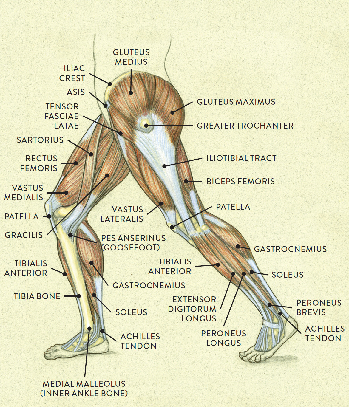
They are associated with muscles discussed in the section above (see. The primary function of the knee is to hinge at the lower extremity. • muscle tissues develop from embryonic cells. Learn about the muscles, tendons, bones, and ligaments that comprise the knee joint anatomy. This is a table of skeletal muscles of the human anatomy.
Muscular System - Muscles of the Human Body from www.innerbody.com You can click the links in the image, or the links below the image to find out more information on any muscle group. How to study muscle anatomy. In this section, learn more about the anatomy of the muscles of the neck. Convergent muscles contain fibers that have a wide origin, but converge in order to attach to a narrow tendon. Inflammation of this region caused by repetitive stress or trauma may lead to pain and numbness known as carpal tunnel syndrome. Attached to the bones of the skeletal system are about 700 named muscles that make up roughly half of a person's body weight. Learning to draw muscles may conjure medical charts in daunting details, but such complexity is unnecessary. The three scalene muscles are found forming the floor of the posterior triangle.
You can click the links in the image, or the links below the image to find out more information on any muscle group.
It elevates and protrudes the mandible. • definitions • introduction • development of muscles • classification • anatomy of skeletal muscle • muscle physiology • properties • muscles of development of muscles. Convergent muscles contain fibers that have a wide origin, but converge in order to attach to a narrow tendon. Skeletal muscles allow the body to move and maintain posture; An interactive tutorial teaching the position, actions, innervation and attachments of the rectus femoris muscle with the aid of anatomical illustrations. Through a simple and intuitive interface it is possible to observe systems: Knee function is determined in large part by the anatomy of the joint. Find the best weight lifting exercises that target each muscle or groups of muscles. Learn about the muscles, tendons, bones, and ligaments that comprise the knee joint anatomy. Anatomy of a muscle cell. Inflammation of this region caused by repetitive stress or trauma may lead to pain and numbness known as carpal tunnel syndrome. The anterior and middle scalenes originate from the transverse processes of certain cervical vertebrae and attach to the first rib. Anatomy of the short head of the biceps brachii muscle.
Each of these muscles is a discrete organ constructed of skeletal muscle tissue, blood vessels, tendons, and nerves. This article provides an overview of the neck muscles, their anatomy, origins, insertions, actions, and innervation. Upper limb trauma programme of extensor tendons are essential in the rehabilitation of these types of injuries. The tendons of these muscles pass through a small corridor in the wrist known as the carpal tunnel. They are associated with muscles discussed in the section above (see.
Lateral view of a pair of legs from schoolbag.info In the diagrams below, i'll be showing muscle groups in color, with a black line to show the forms that would show through the skin (i also show protruding bones that would do the same). This article provides an overview of the neck muscles, their anatomy, origins, insertions, actions, and innervation. In this section, learn more about the anatomy of the muscles of the neck. Want to learn more about it? The tendons of these muscles pass through a small corridor in the wrist known as the carpal tunnel. How to study muscle anatomy. This handbook of general anatomy has been written to meet the requirements of students who are newly admitted to medica. By contracting, they also aid the venous return of blood to the heart and with age, these components of the musculoskeletal system progressively degenerate, which contributes to frailty and increases the risk of falls and fractures.
There's no strict demarcation or dividing line between the tendon and the covering around this muscle but that covering is called is called the epimysium fp my cm and it's really just connective tissue that covers the muscle kind of protects it reduces friction.
Inflammation of this region caused by repetitive stress or trauma may lead to pain and numbness known as carpal tunnel syndrome. This handbook of general anatomy has been written to meet the requirements of students who are newly admitted to medica. It elevates and protrudes the mandible. How to study muscle anatomy. Musculoskeletal, cardiovascular, nervous, respiratory, digestive, urogenital (male and female), endocrine, lymphatic, eye and ear. The muscles of the torso, examined in the previous chapter, include a few that attach directly into the upper arm and help move the humerus at the shoulder joint. Along with lateral pterygoid muscle it produces side to side movement of mandible. Skeletal muscles allow the body to move and maintain posture; Smooth muscle contractions are involuntary movements triggered by. Attached to the bones of the skeletal system are about 700 named muscles that make up roughly half. Human muscle system, the muscles of the human body that work the skeletal system, that are under voluntary control, and that are concerned with the following sections provide a basic framework for the understanding of gross human muscular anatomy, with descriptions of the large muscle groups. Each of these muscles is a discrete organ constructed of skeletal muscle tissue, blood vessels, tendons, and nerves. Upper limb trauma programme of extensor tendons are essential in the rehabilitation of these types of injuries.
Attached to the bones of the skeletal system are about 700 named muscles that make up roughly half. Muscular contraction is necessary for voluntary and involuntary movement of limbs, stabilization of joints, maintaining luminal diameter (in the case of arteries, bowel, etc), and to produce heat. It elevates and protrudes the mandible. Each of these muscles is a discrete organ constructed of skeletal muscle tissue, blood vessels, tendons, and nerves. This is a table of skeletal muscles of the human anatomy.
DIAGRAMS: Arm Muscles Diagram from 2.bp.blogspot.com Learning to draw muscles may conjure medical charts in daunting details, but such complexity is unnecessary. The three scalene muscles are found forming the floor of the posterior triangle. Convergent muscles contain fibers that have a wide origin, but converge in order to attach to a narrow tendon. By contracting, they also aid the venous return of blood to the heart and with age, these components of the musculoskeletal system progressively degenerate, which contributes to frailty and increases the risk of falls and fractures. Find the best weight lifting exercises that target each muscle or groups of muscles. Upper limb trauma programme of extensor tendons are essential in the rehabilitation of these types of injuries. • muscle tissues develop from embryonic cells. How to study muscle anatomy.
Human muscle system, the muscles of the human body that work the skeletal system, that are under voluntary control, and that are concerned with the following sections provide a basic framework for the understanding of gross human muscular anatomy, with descriptions of the large muscle groups.
Knee function is determined in large part by the anatomy of the joint. Inflammation of this region caused by repetitive stress or trauma may lead to pain and numbness known as carpal tunnel syndrome. Topographically, the muscles in this group are classed along with the lateral torso wall and upper extremity, which is due to their location as well as their genetic development based on their embryological origin. Learn about the muscles, tendons, bones, and ligaments that comprise the knee joint anatomy. Muscles of mastication are classified as main and accessory muscles. The muscles of the torso, examined in the previous chapter, include a few that attach directly into the upper arm and help move the humerus at the shoulder joint. Anatomy 3d atlas allows you to study human anatomy in an easy and interactive way. An interactive tutorial teaching the position, actions, innervation and attachments of the rectus femoris muscle with the aid of anatomical illustrations. The anterior and middle scalenes originate from the transverse processes of certain cervical vertebrae and attach to the first rib. Movement of the mandible at the temporomandibular joint). Learning to draw muscles may conjure medical charts in daunting details, but such complexity is unnecessary. • muscle tissues develop from embryonic cells. Learn about human anatomy muscles with free interactive flashcards.